File:Periodontium.png
Archivo original (339 × 665 píxeles; tamaño de archivo: 128 kB; tipo MIME: image/png)
Summary
Download original file 339 × 665 px svg View in browser You need to attribute the author
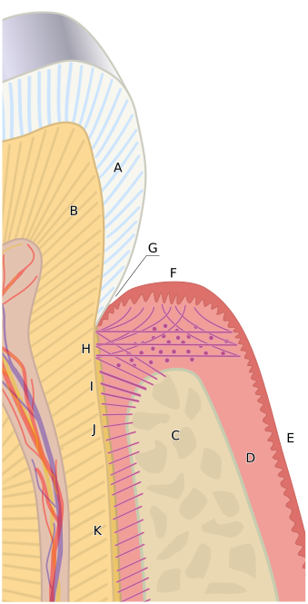
By Goran tek-en, CC BY-SA 4.0, https://commons.wikimedia.org/w/index.php?curid=30709759 A diagram of the periodontium. The crown of the tooth is covered by enamel (A). Dentin (B). The root of the tooth is covered by cementum. C, alveolar bone. D, subepithelial connective tissue. E, oral epithelium. F, free gingival margin. G, gingival sulcus. H, principal gingival fibers. I, alveolar crest fibers of the periodontal ligament (PDL). J, horizontal fibers of the PDL. K, oblique fibers of the PDL. Goran tek-en The Periodontium
CC BY-SA 4.0 File:Periodontium.svg Created: 20 January 2014 About this interface | Discussion | Help
Historial del archivo
Haz clic sobre una fecha y hora para ver el archivo tal como apareció en ese momento.
| Fecha y hora | Miniatura | Dimensiones | Usuario | Comentario | |
|---|---|---|---|---|---|
| actual | 20:43 2 dic 2021 | | 339 × 665 (128 kB) | Rossdonaldson1 (discusión | contribs.) | Download original file 339 × 665 px svg View in browser You need to attribute the author By Goran tek-en, CC BY-SA 4.0, https://commons.wikimedia.org/w/index.php?curid=30709759 A diagram of the periodontium. The crown of the tooth is covered by ename... |
No puedes sobrescribir este archivo.
Usos del archivo
Las siguientes páginas usan este archivo: