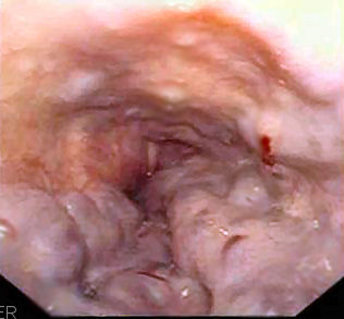
File:Esophageal varices - wale.jpg
No disponible a mayor resolución.
Esophageal_varices_-_wale.jpg (316 × 293 píxeles; tamaño de archivo: 17 kB; tipo MIME: image/jpeg)


Historial del archivo
Haz clic sobre una fecha y hora para ver el archivo tal como apareció en ese momento.
| Fecha y hora | Miniatura | Dimensiones | Usuario | Comentario | |
|---|---|---|---|---|---|
| actual | 21:39 1 nov 2023 | | 316 × 293 (17 kB) | Rossdonaldson1 (discusión | contribs.) | thumb|Posterior view of the position and relation of the esophagus in the cervical region and in the posterior mediastinum. thumb|Layers of the GI track: the mucosa, submucosa, muscularis, and serosa. thumb|Esophagus anatomy and nomenclature based on two systems. |
No puedes sobrescribir este archivo.
Usos del archivo
Las siguientes páginas usan este archivo: