Diferencia entre revisiones de «Hip dislocation»
Sin resumen de edición |
Sin resumen de edición |
||
| (No se muestran 53 ediciones intermedias de 12 usuarios) | |||
| Línea 1: | Línea 1: | ||
==Background== | ==Background== | ||
{{Hip anatomy background images}} | |||
*Orthopedic emergency | |||
**Reduction of native hip should occur within 6hr due to high risk of avascular necrosis | |||
**Hip prosthetic dislocation is more common and less emergent | |||
*High-energy trauma is primary mechanism for native hip dislocation | |||
**Dashboard impact, fall from height, sports injury | |||
*Low-energy trauma can cause hip prosthetic dislocation | |||
**Tying shoes, sitting on toilet or low seat | |||
===Types=== | |||
*Posterior | |||
**90% of hip dislocations | |||
**Often associated with acetabular fracture | |||
*Anterior | |||
**10% of hip dislocations<ref>Holt GE and McCarty EC. Anterior hip dislocation with an associated vascular injury requiring amputation. J Trauma. 2003; 55(1):135-138.</ref> | |||
**Can be superior (pelvic) or inferior (obturator) | |||
**Neurovascular compromise is unusual | |||
=== | ==Clinical Features== | ||
===Posterior Dislocation=== | |||
*Extremity is shortened, internally rotated, adducted | |||
*Neurovascular exam may review sciatic nerve compromise | |||
===Anterior Dislocation=== | |||
*Extremity is extended (superior) or flexed (inferior), externally rotated, abducted<ref>Alonso JE, et al. A review of the treatment of hip dislocations associated with acetabular fractures. Clin Orthop Relat Res. 2000; 377(8):32-43.</ref> | |||
==Differential Diagnosis== | |||
{{Hip pain DDX}} | |||
==Evaluation== | |||
===Workup=== | |||
[[File:HipdisX.png|thumb]] | |||
[[File:Posthipdislocation.jpg|thumb|Post-surgical hip dislocation]] | |||
*Hip AP and lateral views | |||
**Posterior Dislocation: AP view femoral head posterior and superior to acetabulum | |||
**Anterior Dislocation: AP view shows femoral head in obturator foramen (inferior to acetabulum) | |||
**If associated femoral neck fracture, will likely need orthopedics | |||
*Consider Judet views | |||
*Consider knee xray | |||
*Consider CT to evaluate acetabulum for subtle fractures (esp for posterior dislocation) | |||
===Diagnosis=== | |||
*Diagnosed typically via radiograph (see above) | |||
==Management== | |||
*Reduction recommended within 6 hours to prevent avascular necrosis of the femoral head<ref>Jaskulka RA, et al. Dislocation and fracture-dislocation of the hip. J Bone Joint Surg Br. 1991; 73(3):465-469.</ref> | |||
*'''Femoral neck fracture is a contraindication to closed reduction''' | |||
*[[Procedural sedation]] | |||
== | ===Posterior=== | ||
====Allis Maneuver==== | |||
[[File:Hip_Reduction.jpg|thumb|Allis maneuver.]] | |||
*Supine patient on table: deeper sedation ([[propofol]] helps with tissue relaxation); firm distal traction at flexed knee to pull head back into acetabulum; assistant stabilizes pelvis by pushing on ASISs | |||
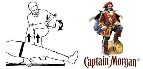
====Captain Morgan Hip Reduction<ref>Hendey GW and Avila AA. The Captain Morgan Technique for the Reduction of the Dislocated Hip. Annals of Emergency Medicine, Volume 60, Issue 1, July 2012, Pages 135-136.</ref>==== | |||
*See figure [http://67.media.tumblr.com/tumblr_lriey37Dpa1qafl51o1_500.png here] | |||
*See video [https://www.youtube.com/watch?v=iCxRMj6h3So here] | |||
*Provider's knee behind supine patients flexed knee with anterior force lifting (via provider plantar flexing foot) and rotation as needed | |||
*Successful in patients with prosthetic hips as well | |||
*Poses less risk of knee injury since most force is applied by lifting leg rather than applying leverage at knee | |||
*Less risk to provider who does not have to stand on top of gurney, and requires only one provider | |||
====The Waddell Technique<ref>Waddell BS, Mohamed S, Glomset JT, Meyer MS. A Detailed Review of Hip Reduction Maneuvers: A Focus on Physician Safety and Introduction of the Waddell Technique. Orthop Rev (Pavia). 2016;8(1):6253.</ref>==== | |||
[[File:Waddell_Technique.jpg|thumb|The Waddell technique]] | |||
*A modified Allis Maneuver that allows the provider to follow back safety recommendations provided by OSHA | |||
*Provider hovers over patient on the bed and places their forearm under the patient's knee | |||
*The provider squats down, draping their forearm over their knees with the elbow on one knee and wrist/hand over the other knee | |||
*Provider then leans back, pivoting on feet and holding the patient's leg close to their chest, while an assistant stabilizes the pelvis | |||
== | ===Anterior=== | ||
*Reduction: traction, internal rotation, and then external rotation once the femoral hip clears the acetabular rim | |||
==Disposition== | |||
*If reduced, outpatient with ortho follow up | |||
===Post Reduction Care=== | |||
*Maintain dislocation precautions: | |||
**Do not bend the operated hip past 90 degrees | |||
***Zimmer splint or other knee immobilizer can help with this as most individuals cannot flex hip without flexing knee | |||
**Do not cross the midline of the body with operated leg (use hip abduction pillow) | |||
**Do not rotate the operated leg inward | |||
**In bed, toes and knee cap should point toward ceiling | |||
*Toe-touch or feather weight-bearing | |||
== | ==Complications== | ||
*Post-traumatic arthritis | |||
**20% in simple dislocations | |||
**Common in complex dislocations | |||
*Femoral head osteonecrosis | |||
**5-40% | |||
**Delay in reduction >6 hours increases risk | |||
*Sciatic nerve injury (check EHL function - toe extension) | |||
**8-20% incidence | |||
**Delay in reduction increases risk | |||
*Recurrent dislocations: <2% | |||
==External Links== | |||
==References== | |||
<references/> | |||
[[Category:Orthopedics]] | |||
[[Category: | |||
Revisión actual - 20:25 26 feb 2025
Background


- Orthopedic emergency
- Reduction of native hip should occur within 6hr due to high risk of avascular necrosis
- Hip prosthetic dislocation is more common and less emergent
- High-energy trauma is primary mechanism for native hip dislocation
- Dashboard impact, fall from height, sports injury
- Low-energy trauma can cause hip prosthetic dislocation
- Tying shoes, sitting on toilet or low seat
Types
- Posterior
- 90% of hip dislocations
- Often associated with acetabular fracture
- Anterior
- 10% of hip dislocations[1]
- Can be superior (pelvic) or inferior (obturator)
- Neurovascular compromise is unusual
Clinical Features
Posterior Dislocation
- Extremity is shortened, internally rotated, adducted
- Neurovascular exam may review sciatic nerve compromise
Anterior Dislocation
- Extremity is extended (superior) or flexed (inferior), externally rotated, abducted[2]
Differential Diagnosis
Hip pain
Acute Trauma
- Femur fracture
- Proximal
- Intracapsular
- Extracapsular
- Shaft
- Mid-shaft femur fracture (all subtrochanteric)
- Proximal
- Hip dislocation
- Pelvic fractures
Chronic/Atraumatic
- Hip bursitis
- Psoas abscess
- Piriformis syndrome
- Meralgia paresthetica
- Septic arthritis
- Obturator nerve entrapment
- Avascular necrosis of hip
Evaluation
Workup


- Hip AP and lateral views
- Posterior Dislocation: AP view femoral head posterior and superior to acetabulum
- Anterior Dislocation: AP view shows femoral head in obturator foramen (inferior to acetabulum)
- If associated femoral neck fracture, will likely need orthopedics
- Consider Judet views
- Consider knee xray
- Consider CT to evaluate acetabulum for subtle fractures (esp for posterior dislocation)
Diagnosis
- Diagnosed typically via radiograph (see above)
Management
- Reduction recommended within 6 hours to prevent avascular necrosis of the femoral head[3]
- Femoral neck fracture is a contraindication to closed reduction
- Procedural sedation
Posterior
Allis Maneuver

- Supine patient on table: deeper sedation (propofol helps with tissue relaxation); firm distal traction at flexed knee to pull head back into acetabulum; assistant stabilizes pelvis by pushing on ASISs
Captain Morgan Hip Reduction[4]
- See figure here
- See video here
- Provider's knee behind supine patients flexed knee with anterior force lifting (via provider plantar flexing foot) and rotation as needed
- Successful in patients with prosthetic hips as well
- Poses less risk of knee injury since most force is applied by lifting leg rather than applying leverage at knee
- Less risk to provider who does not have to stand on top of gurney, and requires only one provider
The Waddell Technique[5]

- A modified Allis Maneuver that allows the provider to follow back safety recommendations provided by OSHA
- Provider hovers over patient on the bed and places their forearm under the patient's knee
- The provider squats down, draping their forearm over their knees with the elbow on one knee and wrist/hand over the other knee
- Provider then leans back, pivoting on feet and holding the patient's leg close to their chest, while an assistant stabilizes the pelvis
Anterior
- Reduction: traction, internal rotation, and then external rotation once the femoral hip clears the acetabular rim
Disposition
- If reduced, outpatient with ortho follow up
Post Reduction Care
- Maintain dislocation precautions:
- Do not bend the operated hip past 90 degrees
- Zimmer splint or other knee immobilizer can help with this as most individuals cannot flex hip without flexing knee
- Do not cross the midline of the body with operated leg (use hip abduction pillow)
- Do not rotate the operated leg inward
- In bed, toes and knee cap should point toward ceiling
- Do not bend the operated hip past 90 degrees
- Toe-touch or feather weight-bearing
Complications
- Post-traumatic arthritis
- 20% in simple dislocations
- Common in complex dislocations
- Femoral head osteonecrosis
- 5-40%
- Delay in reduction >6 hours increases risk
- Sciatic nerve injury (check EHL function - toe extension)
- 8-20% incidence
- Delay in reduction increases risk
- Recurrent dislocations: <2%
External Links
References
- ↑ Holt GE and McCarty EC. Anterior hip dislocation with an associated vascular injury requiring amputation. J Trauma. 2003; 55(1):135-138.
- ↑ Alonso JE, et al. A review of the treatment of hip dislocations associated with acetabular fractures. Clin Orthop Relat Res. 2000; 377(8):32-43.
- ↑ Jaskulka RA, et al. Dislocation and fracture-dislocation of the hip. J Bone Joint Surg Br. 1991; 73(3):465-469.
- ↑ Hendey GW and Avila AA. The Captain Morgan Technique for the Reduction of the Dislocated Hip. Annals of Emergency Medicine, Volume 60, Issue 1, July 2012, Pages 135-136.
- ↑ Waddell BS, Mohamed S, Glomset JT, Meyer MS. A Detailed Review of Hip Reduction Maneuvers: A Focus on Physician Safety and Introduction of the Waddell Technique. Orthop Rev (Pavia). 2016;8(1):6253.